
Using custom atlases
By default, sf-pediatric will use the Brainnetome atlas for preadolescents1 to perform connectomics analyses.
However, different analyses often require different cortical and subcortical atlases. To support flexible connectomics analyses,
users can provide their own custom atlas for connectome construction. This page covers the requirements and assumptions
behind using custom atlases. Additionally, we included test results
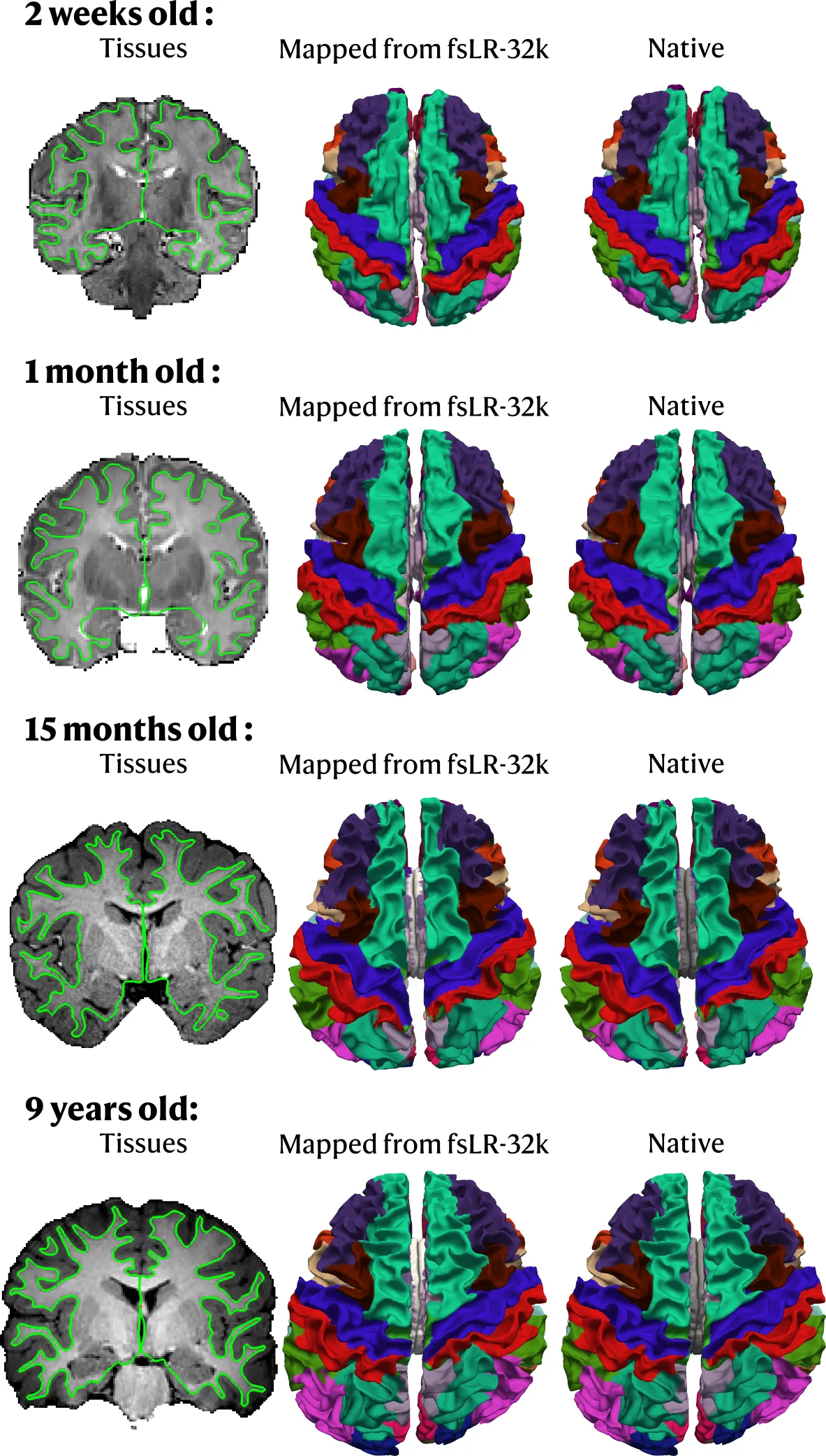
comparing native segmentation with the mapped version from fsLR-32k to subject space.
Requirements
Section titled “Requirements”sf-pediatric relies on surface- and volume-based registration techniques to transform template-space atlases into subject
space. To ensure that mapping between spaces is performed correctly, we follow the BIDS definition for atlases
and require that atlases be in the space-fsLR_den-32k space. Using the default Brainnetome atlas for preadolescents as an
example, the atlas folder should be structured as follows:
Directory<atlases_folder>/BrainnetomeChild/
- atlas-BrainnetomeChild_description.json
Directorytpl-fsLR
- tpl-fsLR_atlas-BrainnetomeChild_den-32k_dseg.dlabel.nii
- tpl-fsLR_atlas-BrainnetomeChild_den-32k_dseg.json
- tpl-fsLR_atlas-BrainnetomeChild_den-32k_dseg.tsv
The top-level atlas-BrainnetomeChild_description.json contains information on how the atlas was derived, relevant references,
the BIDS version, and other metadata. More information can be found here.
The subdirectory tpl-<template> must be named tpl-fsLR, since that is the only currently supported template. It must contain the atlas file
*.dlabel.nii, a corresponding *.json sidecar, and a *.tsv file containing label names, groups, and colors.
As an example, here is the tsv file for the Brainnetome for preadolescents atlas:
| index | label | cifti_label | color_red | color_green | color_blue | opacity |
|---|---|---|---|---|---|---|
| 1 | SFG_L_6_1 | SFG_L_6_1 | 0 | 255 | 0 | 255 |
| 2 | SFG_R_6_1 | SFG_R_6_1 | 0 | 0 | 255 | 255 |
| 3 | SFG_L_6_2 | SFG_L_6_2 | 255 | 0 | 0 | 255 |
| 4 | SFG_R_6_2 | SFG_R_6_2 | 0 | 246 | 255 | 255 |
| … | … | … | … | … | … | … |
The *.dlabel.nii file should contain cortical labels in fsLR-32k space and subcortical structures aligned with the
MNI152NLin6Asym template. To create your atlas in the correct space, we recommend using the Connectome Workbench toolbox.
A few already available atlases can be found in the TemplateFlow archive (be sure that it
contains subcortical structures!).
Use your custom atlas in sf-pediatric
Section titled “Use your custom atlas in sf-pediatric”In order for sf-pediatric to find your custom atlas, specify the folder containing your custom atlas with the
--atlas_folder and the
--atlas_name parameters. By default, sf-pediatric will
look for your atlas at: ${atlas_folder}/${atlas_name}.
In addition to those two parameters, you should also provide the subcortical indexes for your atlas using --subcortical_rois.
This allows the QC report to correctly visualize the segmented subcortical structures. It is highly
recommended that you provide those indexes to get the most out of the QC report.
Example command-line
Section titled “Example command-line”Starting from the basic example described in the a simple example section, specifying
a custom atlas through the params.yml file will look similar to this:
input: "/absolute/path/to/your/BIDS_directory"outdir: "/absolute/path/to/results"atlas_folder: "/absolute/path/to/folder"atlas_name: "BrainnetomeChild"subcortical_rois: "189:224"nextflow run scilus/sf-pediatric -r 0.3.0 \ -params-file params.yml \ -profile docker,tracking \ -resumeHow accurate is the mapping process?
Section titled “How accurate is the mapping process?”Transforming labels between spaces can introduce minor interpolation and registration errors. We evaluated whether mapping from fsLR-32k space to native subject space closely matched native segmentation. Using test data, we performed native segmentation with the Desikan atlas2. For younger participants (< 1 year old), we used a combination of bibsnet3 and infant FreeSurfer4 whereas recon-all-clinical5 was used for older participants. Following native segmentation, we mapped the Desikan atlas (previously transformed into fsLR-32k space and formatted as described in the requirements) from fsLR-32k space to native subject space.
Results are showcased in the figure below. Overall, the mapped and native segmentations showed close agreement across participants and age groups:
References
Section titled “References”1 Li, W., Fan, L., Shi, W., Lu, Y., Li, J., Luo, N., Wang, H., Chu, C., Ma, L., Song, M., Li, K., Cheng, L., Cao, L., & Jiang, T. (2023). Brainnetome atlas of preadolescent children based on anatomical connectivity profiles. Cerebral Cortex, 33(9), 5264–5275. https://doi.org/10.1093/cercor/bhac415
2 Desikan, R. S., Ségonne, F., Fischl, B., Quinn, B. T., Dickerson, B. C., Blacker, D., Buckner, R. L., Dale, A. M., Maguire, R. P., Hyman, B. T., Albert, M. S., & Killiany, R. J. (2006). An automated labeling system for subdividing the human cerebral cortex on MRI scans into gyral based regions of interest. NeuroImage, 31(3), 968–980. https://doi.org/10.1016/j.neuroimage.2006.01.021
3 Hendrickson, T. J., Reiners, P., Moore, L. A., Lundquist, J. T., Fayzullobekova, B., Perrone, A. J., Lee, E. G., Moser, J., Day, T. K. M., Alexopoulos, D., Styner, M., Kardan, O., Chamberlain, T. A., Mummaneni, A., Caldas, H. A., Bower, B., Stoyell, S., Martin, T., Sung, S., … Feczko, E. (2026). BIBSNet: A deep learning baby image brain segmentation network for MRI scans. Developmental Cognitive Neuroscience, 79, 101706. https://doi.org/10.1016/j.dcn.2026.101706
4 Zöllei, L., Iglesias, J. E., Ou, Y., Grant, P. E., & Fischl, B. (2020). Infant FreeSurfer: An automated segmentation and surface extraction pipeline for T1-weighted neuroimaging data of infants 0–2 years. NeuroImage, 218, 116946. https://doi.org/10.1016/j.neuroimage.2020.116946
5 Gopinath, K., Greve, D. N., Magdamo, C., Arnold, S., Das, S., Puonti, O., & Iglesias, J. E. (2025). “Recon-all-clinical”: Cortical surface reconstruction and analysis of heterogeneous clinical brain MRI. Medical Image Analysis, 103, 103608. https://doi.org/10.1016/j.media.2025.103608